10-7-15
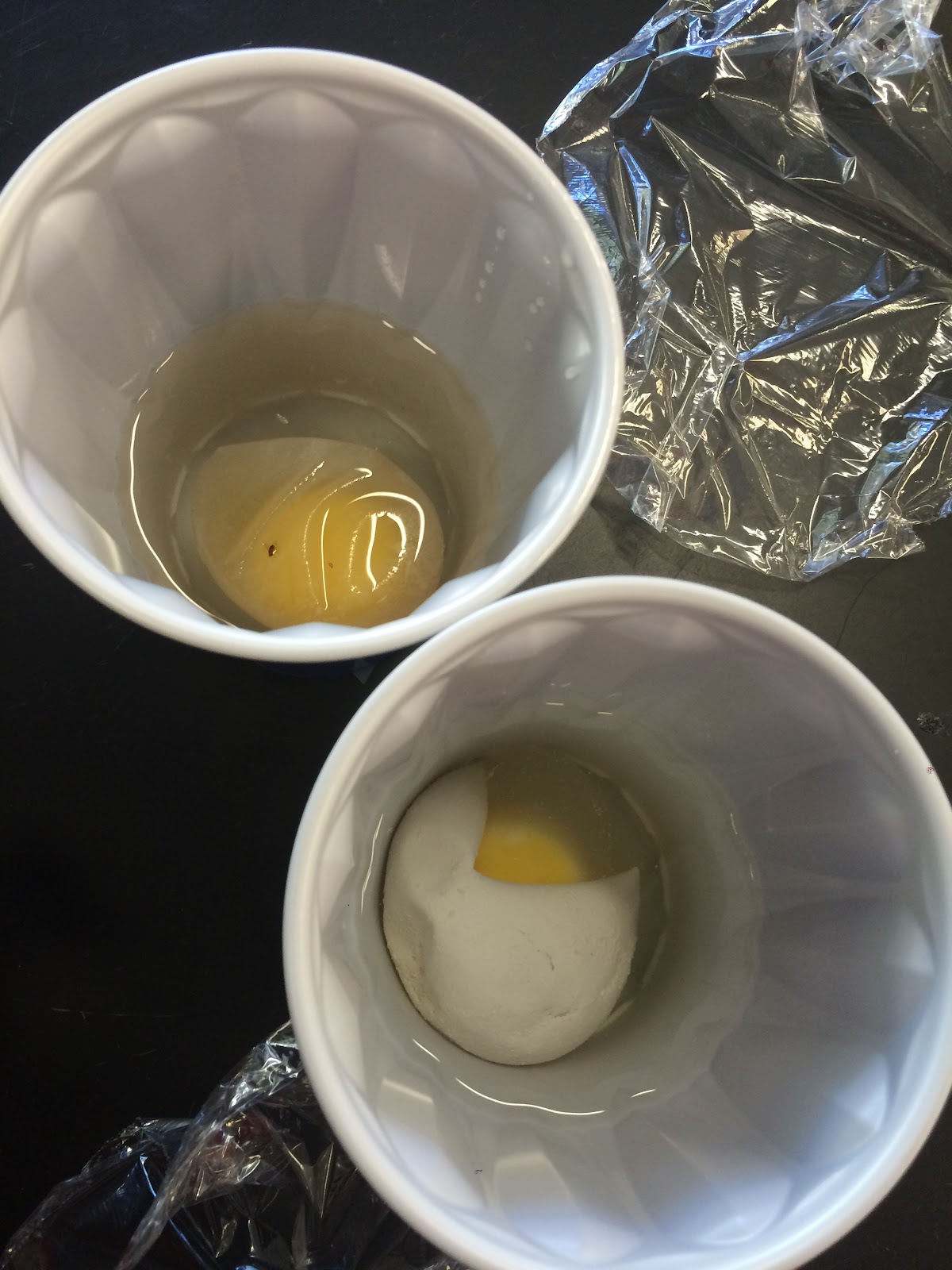
In this lab, we tested eggs to demonstrate how a cell’s internal and external environment changes. First we soaked two eggs in vinegar, which dissolved the egg shell so we could look into the membrane. After measuring their initial circumferences and masses, we placed one egg in deionized water and the other in sugar water. After 2 days we looked at the eggs again and most of the eggs in water burst while the eggs in sugar water shriveled up.
According to the class data, the egg in sugar water became significantly smaller the mass shrinking by about 50% and the circumference shrinking by about 20%. The reason this happened was because the water that was inside the cell went out of it to dilute the sugar that was surrounding the egg. The solvent in this solution was the water and the solute was the sugar.
When a cell’s environment changes it either grows; like in a hypertonic solution, or shrivels up; like in a hypotonic solution. When the egg was in vinegar, it disintegrated the shell and but the egg stayed the same size since it was in an isotonic environment. When the egg was in water it also grew since the water diffused into the egg. While the egg was in the sugar water it shriveled up and shrunk significantly since it sat in a hypotonic solution for so long. The water inside each cell passively diffused across the membrane, from an area of high to low concentration.
This lab mainly helped demonstrate the differences between isotonic, hypotonic, and hypertonic solutions. We experienced an isotonic solution with the vinegar, hypertonic with water, and hypotonic with the sugar.
In life, these applications are used daily by different people. Fresh vegetables are drizzled with water to keep them hydrated and healthy. The water goes inside the cells of the vegetables keeping them big and juicy. Salt is used to de-ice roads because the salts cause the water to diffuse out of the ice, leaving behind a driveable road. But, if there are plants alongside the same road it could do harm to them because the salt will suck out the water it needs to grow and flourish since salt and water is a hypotonic solution.
Based off this experiment I would want to dig deeper to the cellular level and test individual cells under a microscope in these solutions. I would want to see how each organelle is affected rather than a whole egg.
Class Data: Control (DI water) %change
Group#
|
1
|
2
|
3
|
4
|
5
|
6
|
7
|
AVG
|
Mass
|
N/A*
|
N/A*
|
.74
|
.37
|
.45
|
N/A*
|
6.95
|
1.8
|
Circumference
|
N/A*
|
N/A*
|
1.2
|
1.7
|
0
|
N/A*
|
14.37
|
4.3
|
*Egg burst
Class Data: Sugar Water %change
Group#
|
1
|
2
|
3
|
4
|
5
|
6
|
7
| |
Mass
|
-46.70
|
-52.80
|
-52.60
|
-49.70
|
-41.71
|
-39.58
|
-47.70
|
-47.25
|
Circumference
|
-22.40
|
-18.75
|
-26.30
|
-26.60
|
-32.35
|
-21.21
|
-13.00
|
-22.94
|