Sunday, December 6, 2015

DNA Extraction Lab

Conclusion

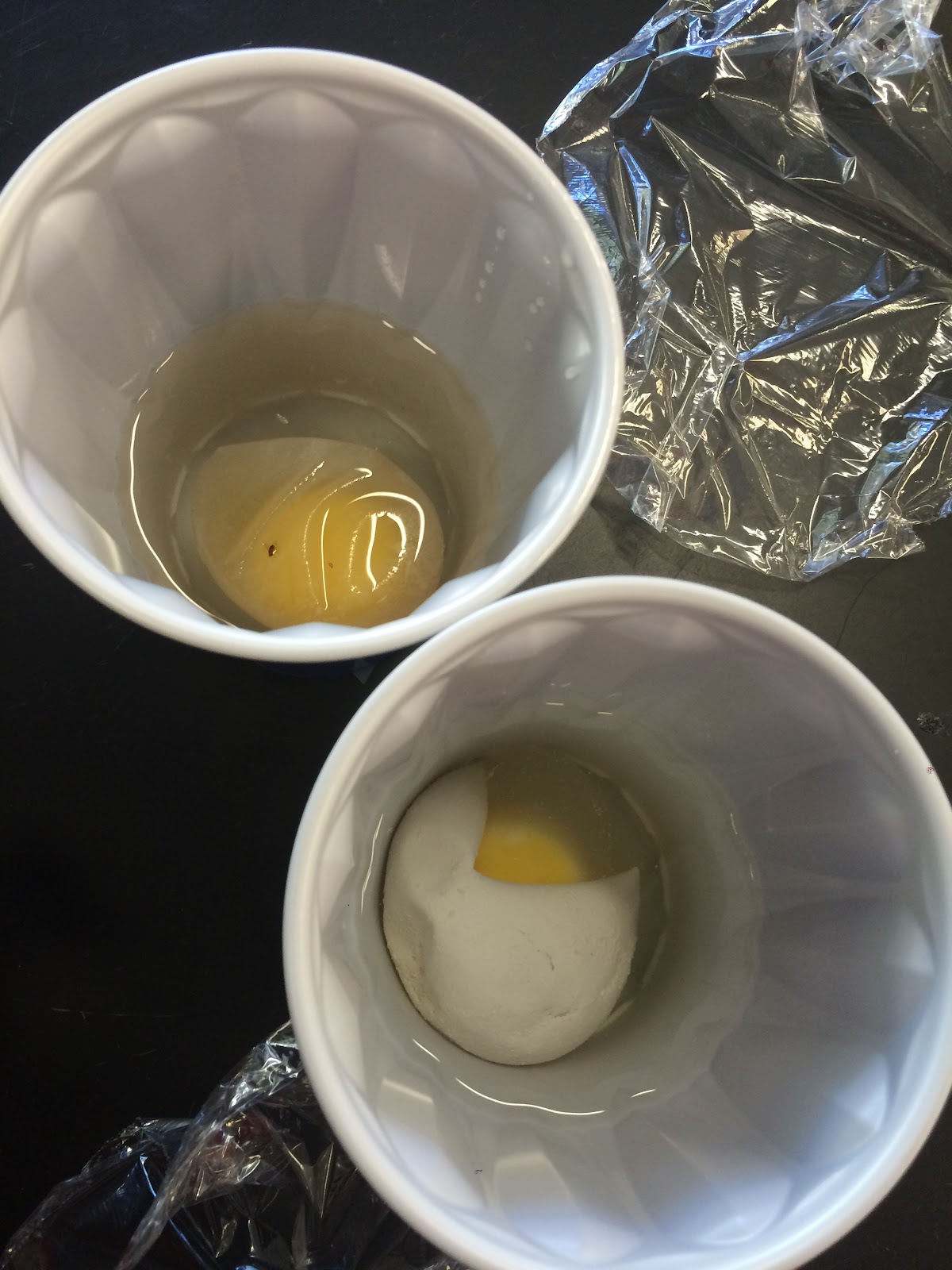
     In this lab we asked the question, "How can DNA be separated from cheek cells in order to study it?" We found that this procedure involved three steps including homogenization, lysis, and precipitation. We carried this out by mixing our cells in a polar liquid. This breaks down the cell and nuclear membranes of the cheek cell, homogenizing it. Afterwards we added soap to the mixture, initiating lysis which disintegrated the membrane. We used pineapple juice to break down histones found in DNA, causing it to uncoil. This was possible because pineapple juice, like a few other liquids, has catabolic proteases and  enzymes, that help to break down the histones. Finally, we poured cold isopropanol alcohol into our test tube and due to its non-polarity combined with the polarity of the DNA, the DNA became a precipitate and rose to the top of the isopropanol alcohol layer.  This data supported our procedure because, to put it simply, it worked!
     While our hypothesis was supported by our data, there was one vital error that we carried out. After adding pineapple juice and soap, we were supposed to gently invert the test tube 6 times and we chose to do this step after adding the alcohol, so the two layers got mixed. Out of the four people in our group, two people's DNA were ruined because of this. This error happened because we created our own procedure without a more educated approval to tell us when to put each step. Another hypothetical error could have been the time factors. If one gargled gatorade for more or less than thirty seconds or observed for more or less than five minutes, results might be changed though the effect isn't drastic. To minimize these rather small to extremely important details, get a teacher's approval before starting and keep a timer handy!
      This lab was done to demonstrate the process of extracting DNA from a cell. From this lab I learnt how to remove DNA from its original position in a cell which helps me understand the concept of DNA and its structure. Based on my experience from this lab I applied the concepts around DNA and I now have a
 sample of my own!


Monday, November 23, 2015

Unit 4 Reflection

This unit was about how things from cells to organisms reproduce. Mitosis and Meiosis along with asexual and sexual reproduction were big concepts and what helped me learn these was the infographic. I understood the process of mitosis so understanding meiosis was quite easy because it was just double the steps with a slightly altered end result. Again, many terms were difficult to memorize but learning them in related bits categorized them in my mind. The infographic helped me study because I went over all the information one last time before taking the test. Check out my Infographic here: https://magic.piktochart.com/output/9417972- biology-infographic Also at the end of the unit, we took a VARK questionare to see what type of learner we are. I got a 12 in kinesthetic and Read/Write learning, and a 10 in both Visual and Aural learner. This surprised me because I thought I was a visual learner but I’m a little bit of everything. That was cool and so was this unit.

Coin Sex Lab Relate and Review

In this lab, we used coins and a partner to simulate the assortment of alleles resulting in different genotypes and phenotypes during sex. The flipping of the coin represented the random combining to create new genes and the probability of a certain type appearing was 50%. The results we got followed the trend of the dihybrid cross simulation but obviously wasn’t exactly the same because real life isn’t probable. We can use probability to state what the possible alleles could be after meiosis or gene segregation, when cells are recombined, and to understand the number of homozygous or heterozygous individuals after a monohybrid cross. In my life, if I had a disease, whether autosomal or X-linked, I could see the possibility of my child going through the same disease by using probability.

Tuesday, November 17, 2015

Genetics Infographic

Below is a picture of my infographic but it is really tiny so if you don't want to kill your vision, click the link! 


Monday, November 2, 2015

Unit 3 Reflection


Unit 3 was about generally about cells. First we learnt of different parts of an animal and plant cell, followed by learning two cell processes, photosynthesis and cellular respiration. This unit was difficult for me because I am really bad at memorizing things and there were a ton of terms. But what helped me was learning the terms as I learnt the processes so I was doing two things that I benefitted from at once. I understood cells in depth during this unit and as a class, it was interesting to dig deeper into specific cell processes. I want to learn more about how plants that live in the ocean where it is hard for sunlight to reach photosynthesize. For the test next week I plan to study by doing the regular reviews with vodcast and textbook notes but this time I will review the questions I got wrong in the CFU quizzes as a new strategy. Hopefully it will help me!

Tuesday, October 27, 2015

Egg Diffusion Lab

10-7-15
In this lab, we tested eggs to demonstrate how a cell’s internal and external environment changes. First we soaked two eggs in vinegar, which dissolved the egg shell so we could look into the membrane. After measuring their initial circumferences and masses, we placed one egg in deionized water and the other in sugar water. After 2 days we looked at the eggs again and most of the eggs in water burst while the eggs in sugar water shriveled up.
According to the class data, the egg in sugar water became significantly smaller the mass shrinking by about 50%  and the circumference shrinking by about 20%. The reason this happened was because the water that was inside the cell went out of it to dilute the sugar that was surrounding the egg. The solvent in this solution was the water and the solute was the sugar.
When a cell’s environment changes it either grows; like in a hypertonic solution, or shrivels up; like in a hypotonic solution. When the egg was in vinegar, it disintegrated the shell and but the egg stayed the same size since it was in an isotonic environment. When the egg was in water it also grew since the water diffused into the egg. While the egg was in the sugar water it shriveled up and shrunk significantly since it sat in a hypotonic solution for so long. The water inside each cell passively diffused across the membrane, from an area of high to low concentration.
This lab mainly helped demonstrate the differences between isotonic, hypotonic, and hypertonic solutions. We experienced an isotonic solution with the vinegar, hypertonic with water, and hypotonic with the sugar.
In life, these applications are used daily by different people. Fresh vegetables are drizzled with water to keep them hydrated and healthy. The water goes inside the cells of the vegetables keeping them big and juicy. Salt is used to de-ice roads because the salts cause the water to diffuse out of the ice, leaving behind a driveable road. But, if there are plants alongside the same road it could do harm to them because the salt will suck out the water it needs to grow and flourish since salt and water is a hypotonic solution.
Based off this experiment I would want to dig deeper to the cellular level and test individual cells under a microscope in these solutions. I would want to see how each organelle is affected rather than a whole egg.
Class Data: Control (DI water)                            %change
Group#
1
2
3
4
5
6
7
AVG
Mass
N/A*
N/A*
.74
.37
.45
N/A*
6.95
1.8
Circumference
N/A*
N/A*
1.2
1.7
0
N/A*
14.37
4.3
*Egg burst

Class Data: Sugar Water                            %change
Group#
1
2
3
4
5
6
7

Mass
-46.70
-52.80
-52.60
-49.70
-41.71
-39.58
-47.70
-47.25
Circumference
-22.40
-18.75
-26.30
-26.60
-32.35
-21.21
-13.00
-22.94

IMG_1978.JPG

Tuesday, October 6, 2015

Egg Macromolecules Lab Conclusions

In this lab we asked the question, “Can macromolecules be identified in an egg cell?” After testing the egg membrane, we found all the macromolecules we tested for, including lipids, proteins, monosaccharides, and polysaccharides. In the yolk we found lipids and the egg white we again contained lipids, proteins, and monosaccharides. On a scale of one to ten, monosaccharides’ existence were a 5 in the membrane and a 2 in the egg white. We knew this because the solution of the egg part and benedicts turned green. Polysaccharides in the membrane were a 7 out of 10 because the color of the iodine mixture turned from light to dark brown. Proteins were a 5 in the membrane and scored a 3 in the egg white because the sodium hydroxide copper sulfate changed from blue to pink/purple. Finally lipids were found in all of the egg, being a 6 in the membrane, 4 in the yolk, and 2 in the egg white. We knew this after the Sudan III color changed from red to  pink/orange. 
One error that may have caused deterred results was the egg not mixing well with the solution used to test its presence. In some directions it said to mix them and in others it didn’t specify. If this happened, some solutions may have a brighter or lighter color and may be interpreted wrong. Another thing that could’ve gone wrong was putting too much or little solution into the egg filled test tubes which could cause too much or too little color, again leading to misinterpreted results. One way to avoid this is to specifically state in the directions the exact amount of drops to put in and whether to stir or not. Another recommendation would be to have more than one test for each egg part just to make sure the results are true. 
This lab was done to model the existence of macromolecules in cells. From this lab I applied the information we learnt in class to real cells which helped me visualize the concepts. By seeing lipids, proteins, and mono/polysaccharides in the cells I took the information from the vodcasts and textbook about macromolecules and related it to cells in eggs and many other areas and how they exist everywhere. Based on my experience from this lab, I was provided with an application of these foundational concepts, which will help me when the class goes deeper into the structure and function of cells.